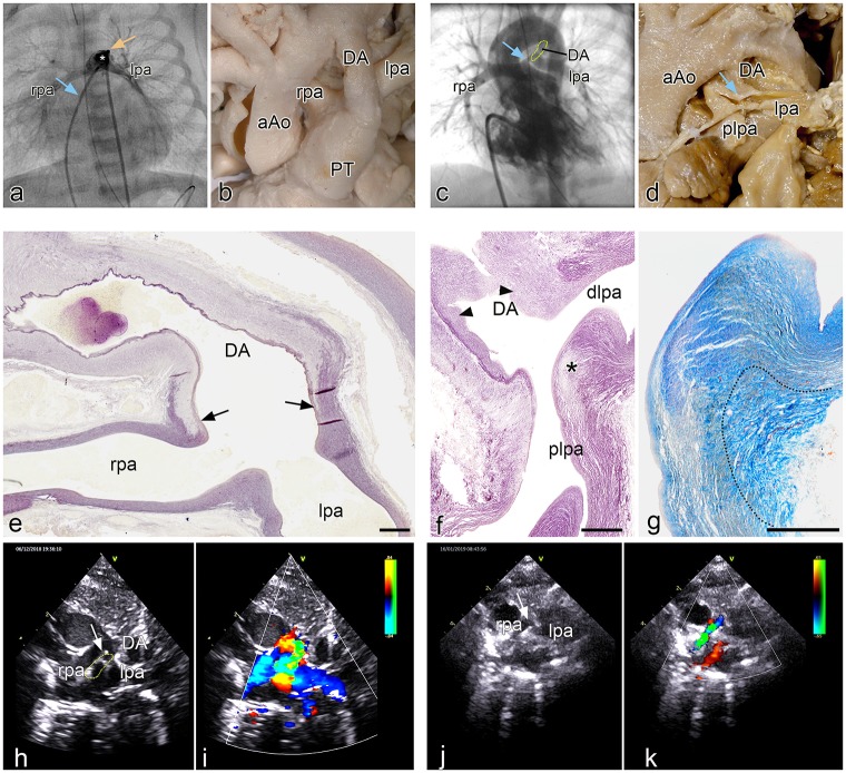
Fig 6.
a. Angio of a patient with pulmonary atresia without VSD in which the leftsided DA (asterisk) is positioned above and anterior of the left pulmonary artery. The left (lpa) and right (rpa) pulmonary arteries (venous line: blue arrow; arterial line yellow arrow) are not compromised at their origin. Fig 6b. Morphology of the arterial pole of a heart specimen in which the ductus arteriosus (DA) connects anteriorly to the pulmonary trunk (PT), while the rpa and lpa are more dorsally connected. Fig 6c. Angio of a patient with tetralogy of Fallot with a marked narrowing (blue arrow) of the proximal (origin) left pulmonary artery (DA dotted area). Fig 6d Postmortem specimen with atresia of the PT and a confluence of the rpa and lpa, with a smaller diameter of the latter. The DA enters sideways into the lpa which shows a PDC at the site of connection (blue arrow). Fig 6e. sagittal section of a human fetal (resorcine fuchsine stained for elastin). The elastin poor muscular DA only lined on the lumen side by an internal elastic lamina, connects with a fishtail like construction (arrows) to the elastin rich rpa and lpa. No DA tissue is encountered in the wall of the lpa and rpa. Fig 6f. Sagittal section (resorcine fuchsin stained for elastin) of a specimen with severe pulmonary ductal coarctation. The tissue of the elastin poor DA, presenting with intimal cushions (arrowheads), is inserted sideways in the wall of the plpa (asterisk), while the dlpa is still elastic in nature. Fig 6g. detail of f in a subsequent section stained for Azan in which it can be seen that the elastic lamellar structure (yellowish) of the dlpa and plpa is interrupted by the adventitia (dotted area) of the DA. Fig 6h. 2D echocardiographic image of the patient with DORV on prostaglandin. In this high parasternal short axis view the rpa, lpa and DA (dotted area) are indicated. The arrow points to the lpa origin. Fig 6i. Same image with Doppler color showing the flow to the rpa and lpa in blue and the DA flow in orange and green. Fig 6j. 2D echo of the same patient 10 days later after placement of the right mBT shunt and discontinuation of prostaglandin. Same view as Fig 6h. The DA has closed. The origin of the lpa is severely stenosed marked by the arrow. Fig 6k. same image as i with showing the flow to the rpa. Because of the mBTS the rpa the flow is turbulent coded in green. The lpa does not receive flow because of the severe stenosis at its origin. The DA has closed. Magnification: e-g bars: 100 μm.

