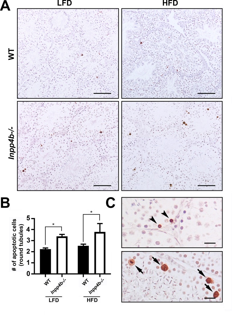
Fig 8. Apoptosis in Inpp4b-/- testis.
A) Increased apoptosis in Inpp4b-/- testes of 3-month old males on LFD and HFD mice. Cell apoptosis was analyzed by TUNEL assay. A representative image from each group is shown. Scale bar represents 200 μm. B) Apoptotic cells (brown) were counted per field under 20X objective in at least 5 fields in each of 2 sections per animal. *p<0.05; n = 3/group. Data shown as mean ± SEM. Statistical analysis was performed using 2-way ANOVA. C) Magnified sections from Inpp4b-/- males on HFD. Spermatogonia and cells in meiotic metaphase showed by arrowheads and arrows respectively. Scale bar is 20 μm.

