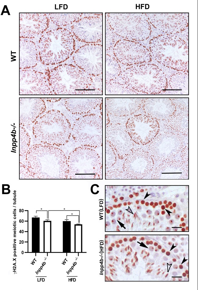
Fig 9. Expression of meiotic marker ϒH2A.X in Inpp4b-/- testis.
A) ϒH2A.X positive cells from the 3-month old WT and Inpp4b-/- males on LFD or HFD. A representative image from each group is shown. Scale bar represents 200 μm. B) The ϒH2A.X-positive cells (brown staining) at prophase I in stage VIII-XI tubules were counted under 40X objective in at least 10 tubules that appear circular on the slide per animal. *p<0.05; n = 3/group. Data shown as mean ± SEM. Statistical analysis was performed using 2-way ANOVA. C) Magnified sections from WT males on LFD and Inpp4b-/- males on HFD, pre-leptone to zygotene spermatocytes, pachytene spermatocytes and elongated spermatids showed by black arrowheads, arrows and white arrowheads respectively. Scale bar is 20 μm.

