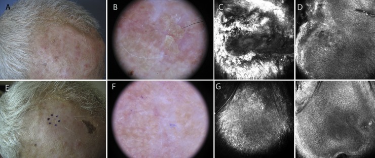
Fig 1. Responder patient treated with ingenol mebutate gel for a grade II actinic keratosis on the forehead (sample AK6).
A: Clinical appearance of the lesion. B: Dermoscopic image showing background erythema and yellow scales with keratotic follicular openings. C: 750×750 μm confocal image at the stratum corneum showing marked hyperkeratosis. D: 750×750 μm confocal image at stratum spinosum demonstrating diffuse atypia of the keratinocytes with different sizes and shapes of the cells. E-F: Clinical and dermoscopic clearance of the lesion after ingenol mebutate treatment. G-H: 750×750 μm confocal images showing normal appearance of the skin.

