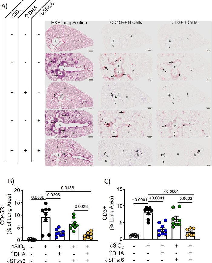
Fig 9. DHA supplementation impedes perivascular and peribronchiolar lymphocyte infiltration, and the neogenesis of ELS.
(A) Light photomicrographs of tissue sections from the left lung of mice intranasally instilled with crystalline silica (cSiO2; +) or saline (VEH alone; -), fed a diet with (+) or without (-) DHA supplementation, and with (+) or without (-) SF.ω6 reduction. Lung sections were stained with H&E (first column), immunohistochemically stained for CD45R+ B lymphoid cells (arrows; brown chromagen; second column), or CD3+ T lymphoid cells (arrows; brown chromagen; third column). Peribronchiolar and perivascular accumulations of B and T lymphoid cells (ELS; arrows) were present in the lungs of cSiO2-exposed mice fed diets without DHA supplementation (second and fourth rows). B or T cell accumulations were not observed in the lung of control mice intranasally instilled with saline and fed CON (row one). Minimal perivascular and peribronchiolar accumulations of B and T lymphoid cells were seen in the lungs of mice intranasally exposed with cSiO2 and fed ↑DHA (row three) or ↓SF.ω6↑DHA. (row five). Abbreviations: a–alveolar parenchyma, b–bronchiolar airway, v–blood vessel. Morphometric analysis was used to quantitatively determine the volume density of (B) CD45R+ and (C) CD3+ area in the measured lung area. Values of p<0.1 are shown, with p<0.05 considered statistically significant.

