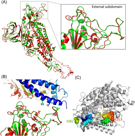
Figure 4.

The binding of receptor‐binding domain (RBD) external subdomain of spike glycoprotein with receptor ACE2. A, Structure comparison of stimulated SARS‐CoV‐2 spike and SARS‐CoV spike glycoprotein (PDB: 5 × 58), SARS‐CoV‐2 spike is shown in red and SARS‐CoV spike is shown in green. RBD contained external subdomain marked in the box. B, The binding model of SARS‐CoV‐2 spike (lower) with human receptor (upper). The possible residues in the interface of SARS‐CoV‐2 spike are shown as sticks. C, Human ACE2 critical for the binding with SARS‐CoV‐2 spike RBD is shown. The key residues located in the interface of ACE2 possibly in combination with spike RBD are shown as a sphere and colored
