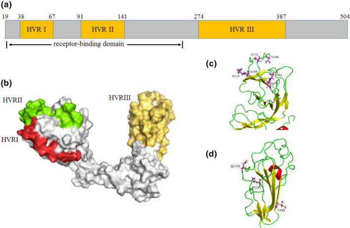
Figure 3.

Characterization and localization of specific nonsynonymous mutations in capsid protein of the newly identified IBV strains in comparison with other IBV isolates. The multi‐alignment of S1 glycoprotein of all IBV genotypes was conducted by Clustal‐Omega, and schematic diagram based on the identified protein functional domains of mature S1 protein (without 1aa‐19aa) was illustrated (a); 3D structure template of GI‐1 genotype (M41 strain) was downloaded from PDB database, and the HVR regions of IBVs (b) and the location of mutation sites of HeN‐2/China/2019 (c and d) were visualized by PyMOL software [Colour figure can be viewed at wileyonlinelibrary.com]
