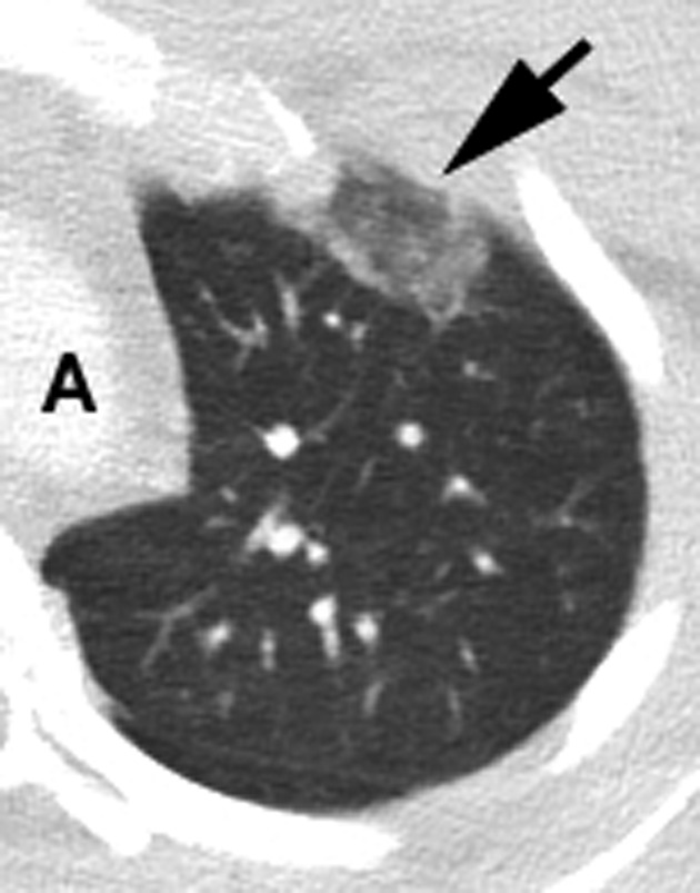
Figure 1.

All images are contrast CT images of the chest in the standard axial plane. For optimal imaging of the lungs, lung windowing was used. All images are from patients admitted during the mid‐weeks of March 2020 to University Medical Center, New Orleans, Louisiana. All patients had documented COVID‐19 by PCR, and respiratory symptoms. Left lung image. A typical GGO is noted (arrow) as a round opacity at the level of the aortic arch, peripherally involving the anterior segment of the left upper lobe. Typically, GGOs are more often found in the basilar location. A = aortic arch
