Figure 2.

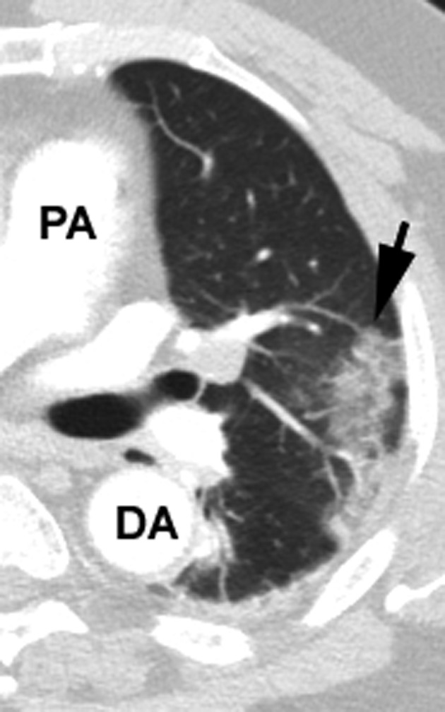
Left lung image. A peripheral GGO (arrow) with reticulation. Reticulation is noted with development of thickening of interlobular septa, appearing as linear opacities. DA = descending aorta; PA = main pulmonary artery

Left lung image. A peripheral GGO (arrow) with reticulation. Reticulation is noted with development of thickening of interlobular septa, appearing as linear opacities. DA = descending aorta; PA = main pulmonary artery