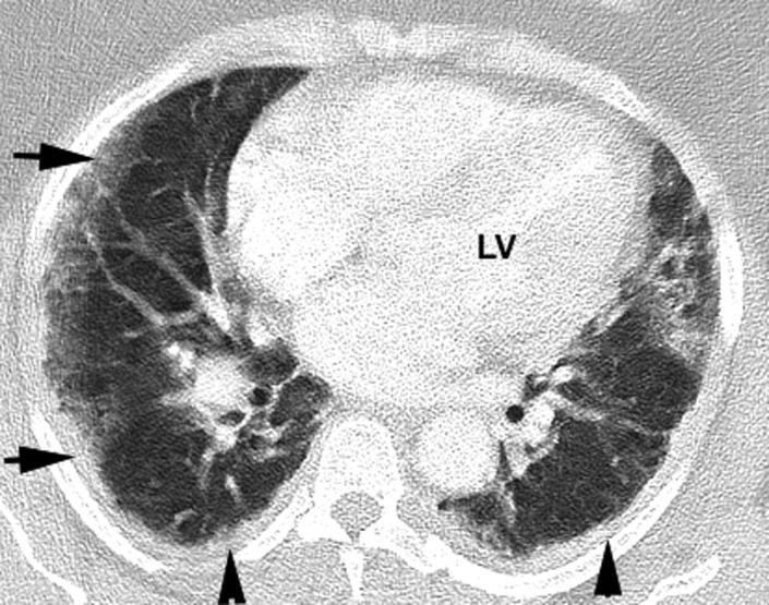
Figure 3.

Level of the heart. Right lung demonstrates an atypical presentation of a peripheral GGO with reticulation, which is not rounded but more band‐like (horizontal arrows). Also, bilateral diffuse band‐like subpleural consolidations are noted (vertical arrows). These band‐like consolidations have been observed in several patients. LV = left ventricle
