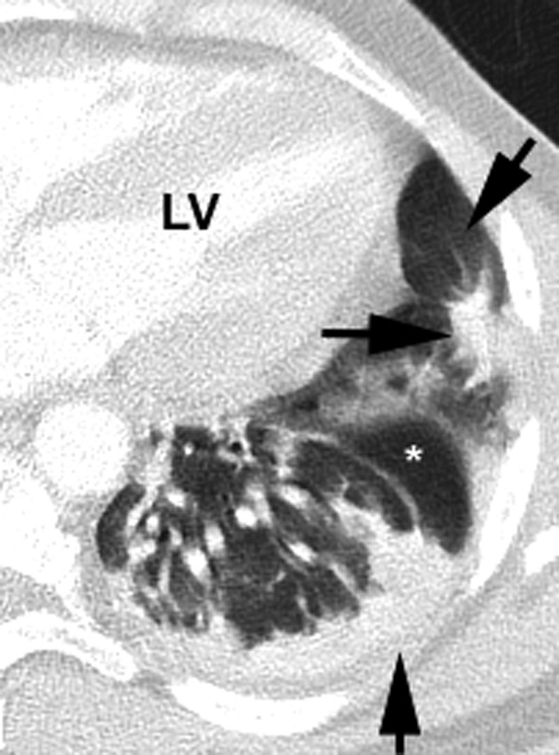
Figure 4.

Left lung image. A zone of GGO (oblique arrow) surrounding an area of consolidation (horizontal arrow). This is termed a halo sign, in which a circular area of GGO is noted around an opacity. An area of normal lung (*) is also noted. As in the prior CT image, a band‐like consolidation is noted (vertical arrow). This is a GGO coalesced into a consolidation with a thick band‐like appearance. LV = left ventricle
