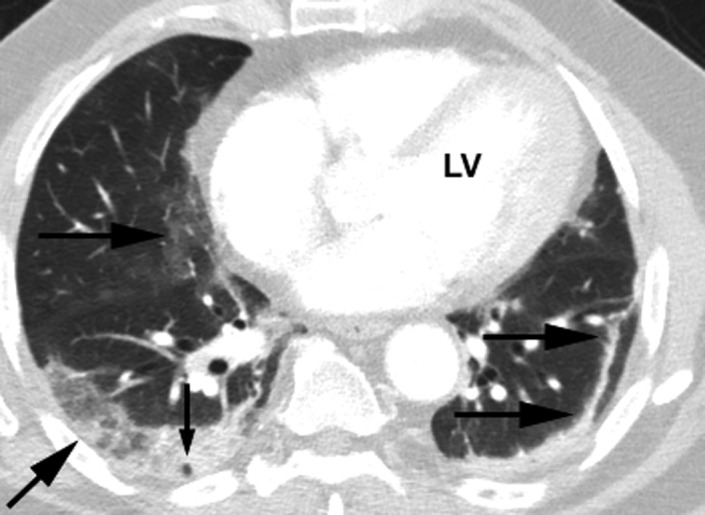
Figure 6.

Right lung ‐ Area of GGO noted more centrally than usually found (horizontal arrow). A peripheral GGO with reticulation (oblique arrow) and the vacuole sign (small vertical arrow). The vacuole sign is a translucent, low‐density shadow within an opacity. Left lung – A parenchymal band (horizontal arrows) is noted. This is defined as a linear opacity, usually up to 3 mm in width and up to 5 cm in length. It may extend to the visceral pleura, which may be retracted where the band attaches. LV = left ventricle
