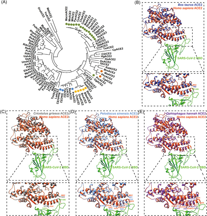
Figure 1.

Structure simulation of SARS‐CoV‐2 RBD with ACE2 from different species. A, Phylogenetic tree of mammalian ACE2. ACE2 proteins from a total of 85 mammals were analyzed by MEGA‐X and the phylogenetic tree was constructed using a maximum‐likelihood method. The green, yellow, orange, and blue represent ACE2 from Primates, Bovidae, Cricetidae, and Cetacea, respectively. B, Structural simulation of the protein complex of Bos taurus ACE2 and SARS‐CoV‐2 RBD. Bos taurus ACE2, Homo sapiens ACE2, and SARS‐CoV‐2 RBD are in medium blue, orange red, and green, respectively. C, Structural simulation of the protein complex of Cricetulus griseus ACE2 and SARS‐CoV‐2 RBD. Cricetulus griseus ACE2, Homo sapiens ACE2, and SARS‐CoV‐2 RBD are in dim gray, orange red, and green, respectively. D, Structural simulation of the protein complex of Pelodiscus sinensis ACE2 and SARS‐CoV‐2 RBD. Pelodiscus sinensis ACE2, Homo sapiens ACE2, and SARS‐CoV‐2 RBD are in cornflower blue, orange red, and green, respectively. E, Structural simulation of the protein complex of Ophiophagus hannah ACE2 and SARS‐CoV‐2 RBD. Ophiophagus hannah ACE2, Homo sapiens ACE2, and SARS‐CoV‐2 RBD are in purple, orange red, and green, respectively. ACE2, angiotensin‐converting enzyme 2; MEGA‐X, Molecular Evolutionary Genetics Analysis version X; RBD, receptor‐binding domain; SARS‐CoV‐2, severe acute respiratory syndrome coronavirus 2
