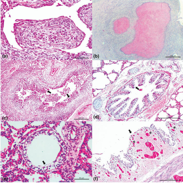
Figure 1.

Histopathologic features observed in dairy cattle with bovine respiratory disease. (a) Observe obliterative bronchiolitis and (b) necrosuppurative bronchopneumonia with large areas of necrosis filled with hypereosinophilic (pink–red) granular debris associated with M. bovis. (c) Observe BVDV associated with necrotizing bronchiolitis (arrows) while (d) ballooning degeneration of bronchial (arrow) (e) and bronchiolar epithelium (arrow) (f) and focal area of necrohemorrhagic bronchitis with angiogenesis (asterisk) at the lamina propria (arrow) with detachment of the bronchial epithelium within the lumen were seen with BoHV‐1. M. bovis‐associated lesions at A and B; BVDV associated lesion at C; and BoHV‐1‐associated lesions at D‐F. Hematoxylin and eosin stain. A, E 50 μm; B‐D, F 200 μm [Colour figure can be viewed at wileyonlinelibrary.com]
