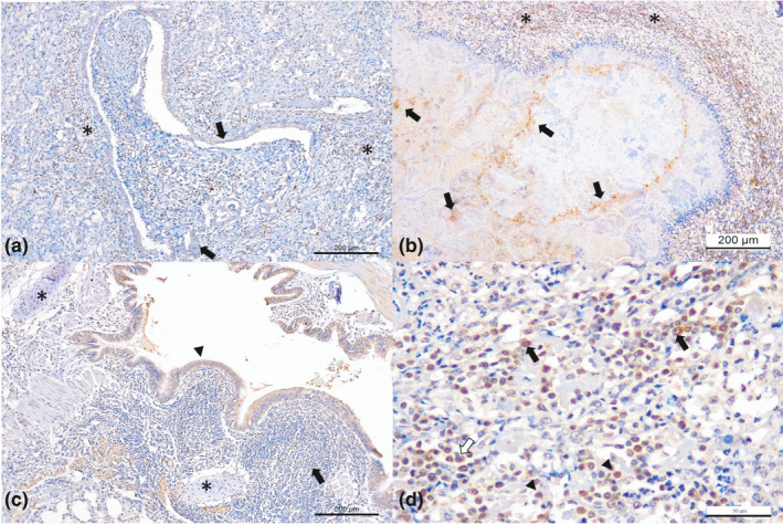
Figure 2.

Immunohistochemical findings observed in dairy cattle with bovine respiratory disease associated with Mycoplasma bovis. (a) There is positive intracytoplasmic immunoreactivity to antigens of M. bovis in obliterative bronchiolitis; observe immunoreactivity within epithelial cells of the bronchiole (arrows) and peripheral macrophage (asterisk). (b) There is necrosuppurative bronchopneumonia with intralesional immunolabelling of bacteria within foci of necrosis (arrows), (c) bronchiolar epithelium (arrow head), at the bronchial hyaline cartilage (asterisk) and peribronchial lymphocytic cuffings (arrow). (d) Closer view of peribronquial lymphocytic cuffing demonstrating positive immunoreactivity for M. bovis with macrophages (black arrows), lymphocytes (arrow head) and plasma cells (red arrow). Immunoperoxidase counterstained with haematoxylin. Bar, A–C 200 μm; D 50 μm [Colour figure can be viewed at wileyonlinelibrary.com]
