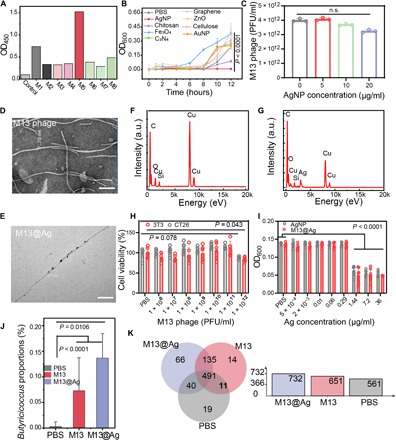
Fig. 3. Characterization of bioinorganic hybridization phages.

(A) In vitro biopanning of specifically Fn-binding M13 phages by Ph.D.-12 peptide phage display library. Eight kinds of clones were examined by ELISA. (B) Screening of a variety of antibacterial nanoparticles (n = 6). (C) Assessment bioactivity of M13 phages after directly assembling with AgNP (n = 3). TEM images of M13 phages (D) and M13@Ag (E), in which M13 phages were native stained. Scale bars, 100 nm. Analysis of element distribution on the surface of M13 phages (F) and M13@Ag (G) detected with EDX. (H) Cell viability of cancerous CT26 cells and normal 3T3 cells after coculture with M13 phages (n = 6). (I) Antibacterial activity of M13@Ag by incubating with Fn for 8 hours (n = 6). (J) Genus-level analysis in the feces from the Fn-colonization CRC murine model with significant increase in antitumor bacteria of Butyricicoccus after PBS, M13, and M13@Ag treatment (n = 3). (K) Venn diagram of identified fecal bacterial strains in Fn-colonization CRC murine model with different treatments (n = 3). Significant differences were assessed in (C) and (J) using one-way ANOVA and t test and in (B), (H), and (I) using t test. The mean values and SD are presented. n.s., not significant; OD, optical density; a.u., arbitrary units.
