. 2020 May 4;9:e56655. doi: 10.7554/eLife.56655

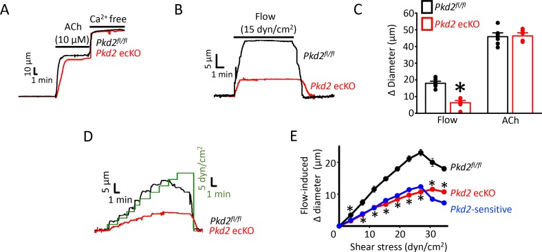
Figure 2. PKD2 channels contribute to intravascular flow-, but not ACh-, mediated vasodilation.

(A) Original traces illustrating responses to ACh (10 µM) and Ca2+-free solution (passive diameter) in pressurized (80 mmHg) mesenteric arteries from Pkd2fl/fl and Pkd2 ecKO mice. (B) Original trace of flow-mediated dilation in pressurized (80 mmHg) mesenteric arteries from Pkd2fl/fl and Pkd2 ecKO mice. (C) Mean diameter changes in response to flow (15 dyn/cm2) or ACh (10 µM). *p<0.05 vs. Pkd2fl/fl. n = 8 for each. # p<0.05 vs. flow in the same genotype. (D) Original traces illustrating diameter responses to stepwise increases in intravascular flow in pressurized (80 mmHg) mesenteric arteries from Pkd2fl/fl and Pkd2 ecKO mice. (E) Mean data. The Pkd2-sensitive component of flow-mediated vasodilation is illustrated in blue. *p<0.05 vs. Pkd2fl/fl. n = 5 for Pkd2fl/fl, n = 4 for Pkd2 ecKO.

Figure 2.

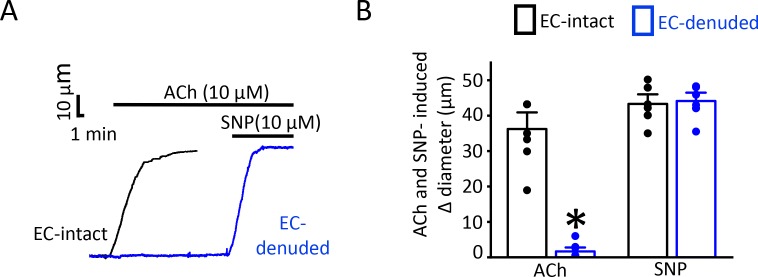
Figure 2—figure supplement 1. Endothelial denudation abolishes ACh-mediated vasodilation.

Figure 2—figure supplement 1.

(A) Original traces demonstrating responses to ACh (10 µM) and SNP (10 µM) in EC-intact (black) and EC-denuded (blue) pressurized (80 mmHg) mesenteric arteries of Pkd2fl/fl. (B) Mean data. *p<0.05 vs. EC-intact. n = 8 for each.
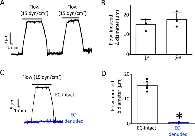
Figure 2—figure supplement 2. Endothelial denudation abolishes flow-mediated dilation.

Figure 2—figure supplement 2.

(A) Original traces demonstrating reproducible flow responses (15 dyn/cm2) in pressurized Pkd2fl/fl mesenteric arteries. (B) Mean diameter changes in response to two consecutive flow (15 dyn/cm2) stimuli. n = 3. (C) Original traces demonstrating flow responses (15 dyn/cm2) in pressurized (80 mmHg) EC-intact and EC-denuded mesenteric arteries from Pkd2fl/fl mice. (D) Mean data for Pkd2fl/fl arteries. n = 7 for EC-intact. n = 6 for Pkd2fl/fl EC-intact. *p<0.05 vs.EC-intact. n = 8 for each.
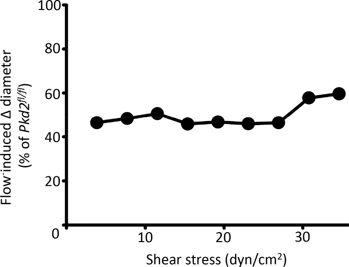
Figure 2—figure supplement 3. Endothelial cell PKD2 knockout attenuates flow-mediated vasodilation over a broad shear stress range.

Figure 2—figure supplement 3.

Mean data illustrating relative dilation to shear stress in pressurized (80 mmHg) Pkd2 ecKO arteries compared with Pkd2fl/fl arteries. n = 4–5.
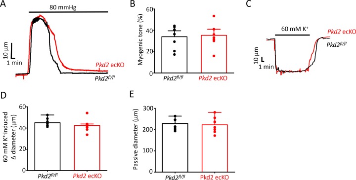
Figure 2—figure supplement 4. Smooth muscle-specific vasoconstriction and passive diameter are unaltered in Pkd2 ecKO arteries.

Figure 2—figure supplement 4.

(A) Representative traces illustrating the development of myogenic tone in pressurized (80 mmHg) Pkd2fl/fl and Pkd2 ecKO arteries. (B) Mean myogenic tone in pressurized (80 mmHg) mesenteric arteries from Pkd2fl/fl and Pkd2 ecKO. n = 8 for each. (C) Representative traces illustrating 60 mm K+ constriction in pressurized (10 mmHg) arteries. (D) Mean data for 60 mM K+-induced constriction. n = 8. (E) Mean data for passive diameter (Ca2+- free PSS) in pressurized (80 mmHg) arteries. n = 8.