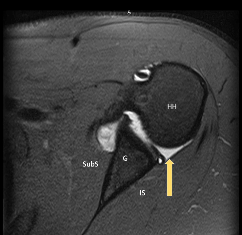
Figure 2. Magnetic Resonance Imaging (MRI) Demonstrating a Posterior Labral Tear.
Axial plane T2 fat-saturated image demonstrating a displaced posterior labral tear in a symptomatic athlete with a batter’s shoulder injury.
Arrow - Posterior labral tear
HH - Humeral head; G - Glenoid; SubS - Subscapularis; IS - Infraspinatus

