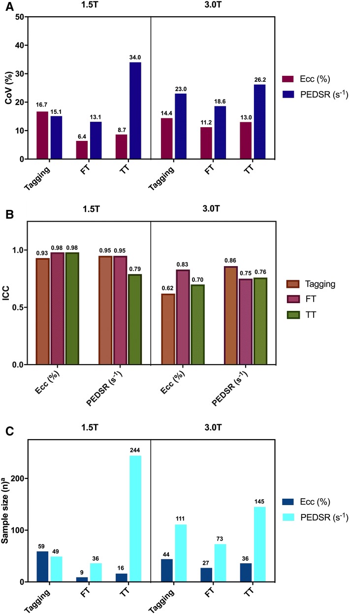
Fig. 4.
Inter-study repeatability of Ecc and PEDSR by tagging, FT and TT at 1.5 T and 3.0 T CMR. Charts showing (a) CoV, (b) ICC and (c) Sample size required to detect a 10% relative change (α = 0.05, Power = 90%). CMR cardiovascular magnetic resonance, CoV coefficient of variation, Ecc global circumferential strain, FT feature tracking, ICC intra-class correlation coefficient, PEDSR global circumferential peak-early diastolic strain rate, TT tissue tracking

