Fig. 1.
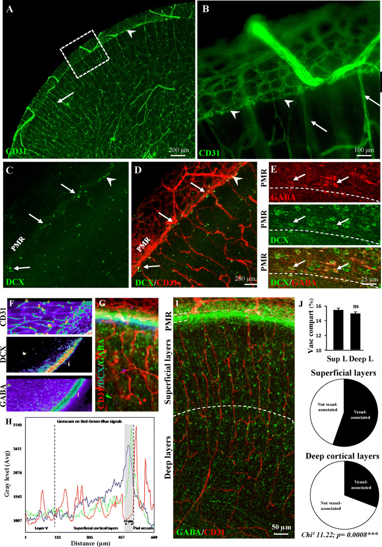
Immunohistochemical characterization of the pial migratory route (PMR) in mouse neonates. a, b Low-magnification (a) and high-magnification (b) photographs visualizing cortical microvessels in transversal cortical slices labeled with CD31 antibodies at P2. Arrow heads indicate a thin network of vessels at the level of the PMR. Arrows indicate cortical radial microvessels. c, d Double immunolabeling experiments showing a low-magnification DCX-positive cells (c) lining the PMR (d; arrowhead). Arrows indicate DCX-positive cells stacking at the level of radial microvessels arising from the PMR. e Double immunolabeling experiments showing at high-magnification tangential cells along the PMR immunoreactive for DCX and GABA (arrows). f–h Line scan analysis (f) of the CD31 (red), DCX (blue) and GABA (green) fluorescent signals acquired from a cultured brain slice at P2 (g). Intensity profiles (h) indicate that DCX and GABA immunofluorescences overlap and border the inner part of pial vessels. i Confocal acquisition of GABA-immunoreactive cells and CD31-labeled microvessels in the developing neocortex at P2. Note a marked vascular interaction of GABA interneurons along radial microvessels. j Quantification of vessel density (upper panel) and vessel-associated GABAergic interneurons in the superficial (middle) and deep (lower panel) cortical layers at P2. Note that the vessel association is preferentially observed in the developing superficial layers. ns not statistically different vs superficial layers. The tests used for the statistical analysis, the number of independent experiments, the number of measures per experiment, and p values are detailed in Table 1

