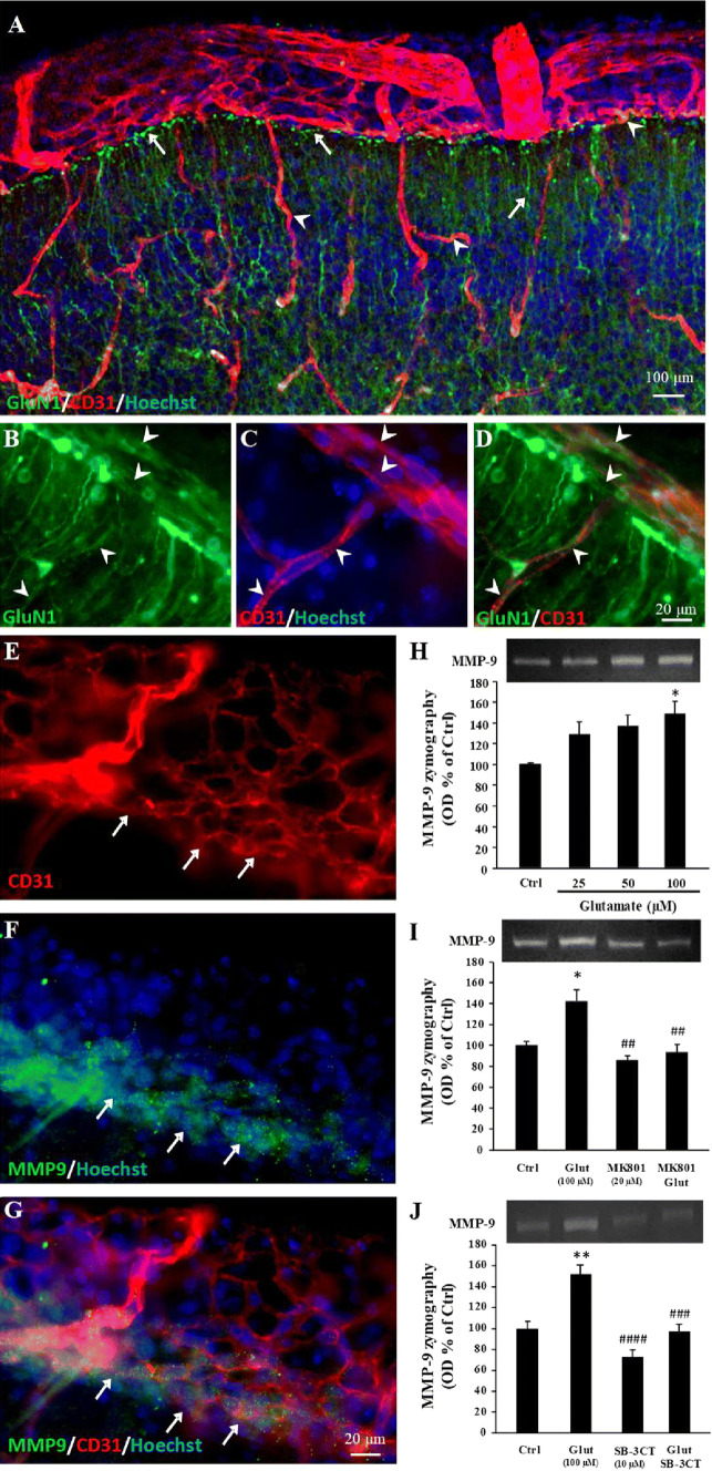
Fig. 2.
GluN1 immunoreactivity in microvessels from the pial migratory route (PMR) and glutamate-dependent regulation of MMP-9 activity. a–d Double immunolabeling experiments showing CD31-positive microvessels and GluN1 immunoreactivity in the neocortex of P2 mice. Immunohistochemistry reveals numerous GluN1-positive cortical radial fibers (a; arrows) and GluN1-positive endothelial cells in the pial migratory route (b–d; arrowheads). e–g Double immunolabeling experiments showing CD31-positive microvessels (e) and MMP-9 immunoreactivity (f) in vessels from the pial migratory route. The overlay (g) indicates that MMP-9 immunoreactivity mainly co-localizes with endothelial cells present in the inner part of the migratory route (arrows). h Quantification by gel zymography of the effects of 6-h exposure of P2 cortical slices to graded concentrations (25–100 µM) of glutamate on MMP-9 activity. i Quantification by gel zymography of the effect of the NMDA-antagonist MK801 (20 µM) on the glutamate-induced increase of MMP-9 activity. j Quantification by gel zymography of the effect of the MMP-9 inhibitor SB-3CT (10 µM) on the glutamate-induced increase of MMP-9 activity. *p < 0.05; **p < 0.01 vs control and ##p < 0.01; ###p < 0.001; ####p < 0.0001 vs glutamate. The tests used for the statistical analysis, the number of independent experiments, the number of measures per experiment, and p values are detailed in Table 1

