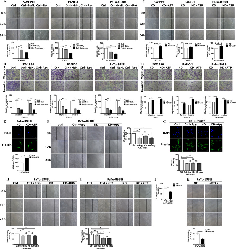
Fig. 5. COX6B2 promotes PDAC cells metastasis via ATP/purinergic receptor pathway.
a, b The migration and invasion ability of SW1990, PANC-1, and PaTu-8988t cells with or without NaN3 (50 μM) or rotenone (200 nM) was evaluated using wound healing assays (a) and trans-well assays (b). c, d The migration and invasion ability of SW1990, PANC-1, and PaTu-8988t cells with knockdown of COX6B2 with or without ATP (100 μM) was evaluated using wound healing assays (c) and transwell assays (d). e Representative immunofluorescence photomicrographs captured using a confocal laser microscope (600× magnification, scale bar = 25 μm), COX6B2 KD PaTu-8988t cells with or without ATP (100 μM) were probed with F-actin (1:100) and a fluorescently labeled IgG-Alexa Fluor 488 secondary antibody (1:500). f Wound healing assays of COX6B2 KD PaTu-8988t cells and paired control cells with or without apyrase (2.5 mU/mL). g Representative immunofluorescence photomicrographs of COX6B2 KD PaTu-8988t cells and paired control cells with or without apyrase (2.5 mU/mL). Cells were probed with anti-F-actin (1:100) and a fluorescently labeled IgG-Alexa Fluor 488 secondary antibody (1:500). Photomicrographs were captured using a confocal laser microscope (600× magnification, scale bar = 25 μm). h, i Wound healing assays of COX6B2 KD PaTu-8988t cells and paired control cells with or without BBG (10 μM) or RB2 (10 μM). j qRT-PCR analysis demonstrates the knockdown efficiency of P2X7 in PaTu-8988t cells. k Wound-healing assays of P2X7 KD PaTu-8988t cells. All data are presented as mean ± SEM (n ≥ 3). *P < 0.05, **P < 0.01, and ***P < 0.001. NS no significance.

