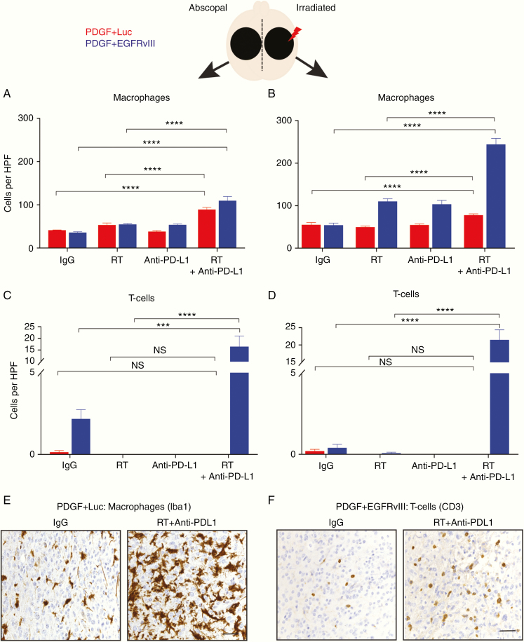
Fig. 3.
Localized radiation and systemic anti–PD-L1 immunotherapy promote intratumoral macrophage and T-cell infiltration. (A, B) Quantification of Iba1+ macrophage influx into unirradiated or abscopal side (A) and radiated side (B) based on IHC on PDGF + Luc (red) and PDGF + EGFRvIII (blue) mouse gliomas on posttreatment day 10, n = 5 mice per group. (C, D) Quantification of CD3+ T-cell macrophage influx into unirradiated or abscopal side (C) and radiated side (D) based on IHC on PDGF + Luc (red) and PDGF + EGFRvIII (blue) mouse gliomas following treatment, n = 5 mice per group. (E, F) Representative IHC for macrophages (Iba1) in PDGF + Luc gliomas (E) and T cells (CD3) in PDGF + EGFRvIII gliomas (F) following treatment (abscopal side). Three to 5 high-powered fields were counted per tumor sample from 5 different tumors on posttreatment day 10. Scale bar = 50 μm. P-values derived from the non-parametric Mann–Whitney U-test, two-sided. Holm–Bonferroni multiple comparison post hoc test for A–D. Error bars = SEM. *P < 0.05, **P < 0.01, ***P < 0.001, ****P < 0.0001, NS = not significant.

