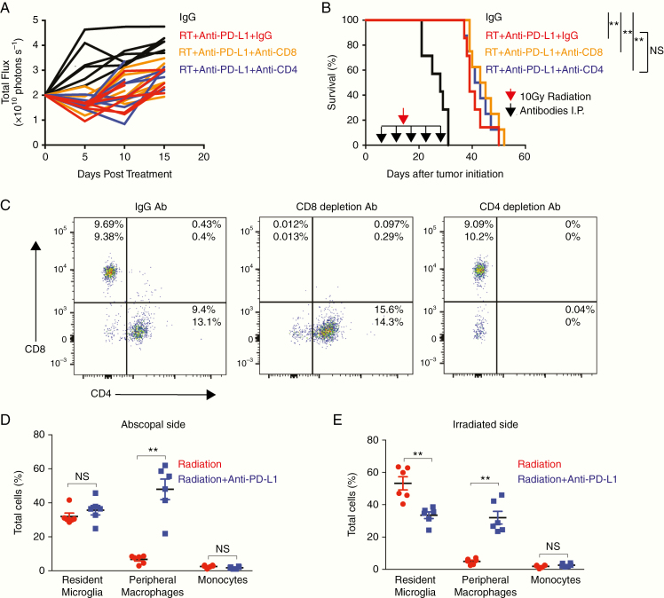
Fig. 4.
T-cell depletion does not affect abscopal effect in PDGF + Luc glioblastoma. (A, B) Bioluminescence (A) and survival data (B) from Tg(NTva);Ink4a-Arf-/-;Pten-/-; LoxP-Stop-LoxP luciferase mice with left-sided PDGF-Pten-/- luciferase tumor and right-sided PDGF-shPten tumor treated with anti-mouse IgG2b isotype control (n = 7), anti-CD8 (n = 8), and anti-CD4 (n = 8) depleting antibodies at 10, 15, 19, 22, and 30 days post-tumor initiation (black arrows in B). Unilateral 10 Gy radiation was then administered (red arrow in B) followed by daily anti–PD-L1 therapy. Based on luciferase activity measured within 10 days post treatment initiation, abscopal response seen in 7/7 IgG treated mice, 8/8 CD8+ T-cell depleted mice, and 8/8 CD4+ T-cell depleted mice. Survival curves following T-cell depletion in radiation and anti–PD-L1 treated mice showing no impact of T-cell depletion on survival (B). Log-rank Mantel–Cox test for radiation alone versus radiation with anti–PD-L1. **P < 0.01, NS = not significant. (C) To verify T-cell depletion, flow cytometry was performed on spleens harvested from mice receiving combination therapy (n = 2 per group) on post-tumor initiation day 22 after 2 doses of depleting antibodies demonstrating >90% depletion. (D, E) On posttreatment day 5, relative to radiation alone (n = 6, red), combination immunotherapy with anti–PD-L1 (n = 6, blue) is associated with increased infiltration of bone marrow–derived macrophages (CX3CR1+/GFP/CCR2+/RFP) into the non-irradiated/abscopal side as shown in D, and the irradiated side as shown in E, of bilateral RCAS-PDGF–driven glioblastoma in Ntva; Cdkn2awt; Ptenflox/flox; CX3CR1+/GFP; CCR2+/RFP mice. Red circles are unilateral radiation treated mice, blue squares are unilateral radiation and anti–PD-L1 treated mice. P-values derived from the non-parametric Mann–Whitney U-test, two-sided. Error bars = SEM. **P < 0.01, NS = not significant.

