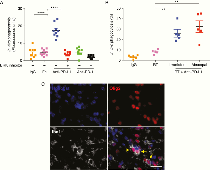
Fig. 6.
Anti–PD-L1 enhances macrophage phagocytosis in PDGF + Luc gliomas. (A) Anti–PD-L1 antibodies promote phagocytosis of fluorescein isothiocyanate–labeled Escherichiacoli particles in vitro via ERK signaling in PD-L1+/PD1− murine macrophages. Phagocytosis of the particles was measured by a Vybrant Phagocytosis Assay Kit. This anti–PD-L1 effect on macrophages is inhibited by PD98059, an ERK inhibitor (20 μM). Three biological replicates each with 3 technical replicates are shown. Orange are IgG controls, pink is FcγR (CD16/32) receptor block, blue is anti–PD-L1 without ERK inhibitor, red is anti–PD-L1 with ERK inhibitor, green is anti-PD1 without ERK inhibitor, black is anti–PD-L1 with ERK inhibitor. (B) Percent phagocytosis represents the proportion of Iba1+ cells (macrophages) that have circumferentially encircled an Olig2+ neoplastic cell. Counts from at least 5 high-powered fields (40x objective) per animal from posttreatment day 10. Each point represents a single animal with bilateral tumors from PDGF + Luc glioblastoma (n = 5–6 mice per treatment group). Orange is IgG control, pink is unilateral radiation only, blue is the irradiated side of anti–PD-L1 with unilateral radiation, red is the abscopal side of anti–PD-L1 with unilateral radiation. (C) Representative immunofluorescence microscopy for Hoechst (all nuclei), Olig2 (neoplastic cells), and Iba1 (macrophages) in PDGF + Luc (radiated side) mouse glioblastoma on posttreatment day 10. Yellow arrows indicate phagocytosed Olig2+ neoplastic cells (magnification 40x; scale bar = 30 μm). P-values derived from the non-parametric Mann–Whitney U-test, two-sided. Error bars = SEM. **P < 0.01, ****P < 0.0001, NS = not significant.

