Fig. 3.
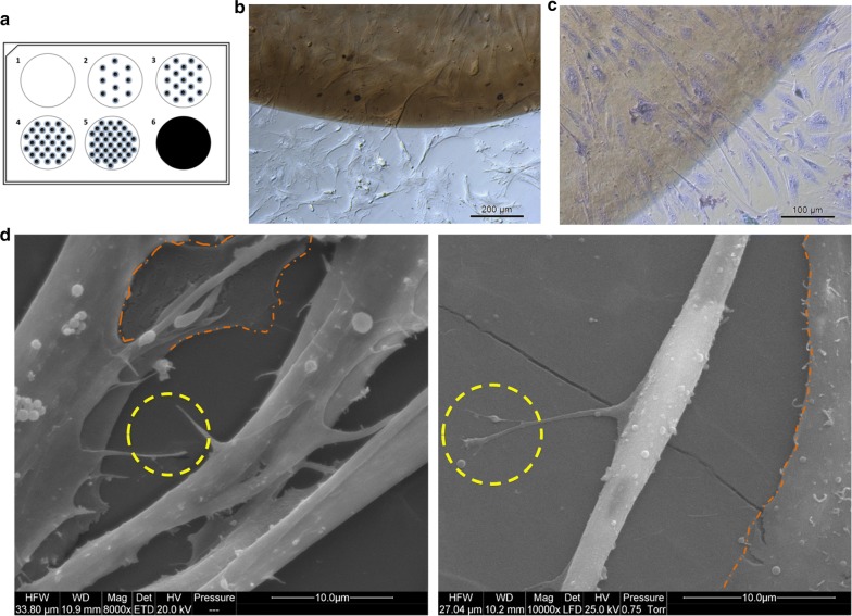
The visualisation of graphene oxide nanifilm (nGO) biocompatibility with mesenchymal stem cells after 48 h of incubation (magnification ×10 and ×20) (b, c) or after 5 days of incubation (c; magnification ×8000 and ×10,000). a The pattern of covering the surface of the culture plate with nGO: Well 1 = control (without nGO); wells 2 to 5 = gradually increasing the number of nGO dots; well 6 = complete surface coverage with nGO. b Live imaging observation of cells migration towards nGO dots. c Hematoxylin and eosin staining of cells growing on nGO dot. d Scanning electron microscopy visualisation of primary muscle fibre grown on nGO; the yellow rings indicate the cell insert directly connected to nGO; the orange surround shows the primary stromal cells base layer

