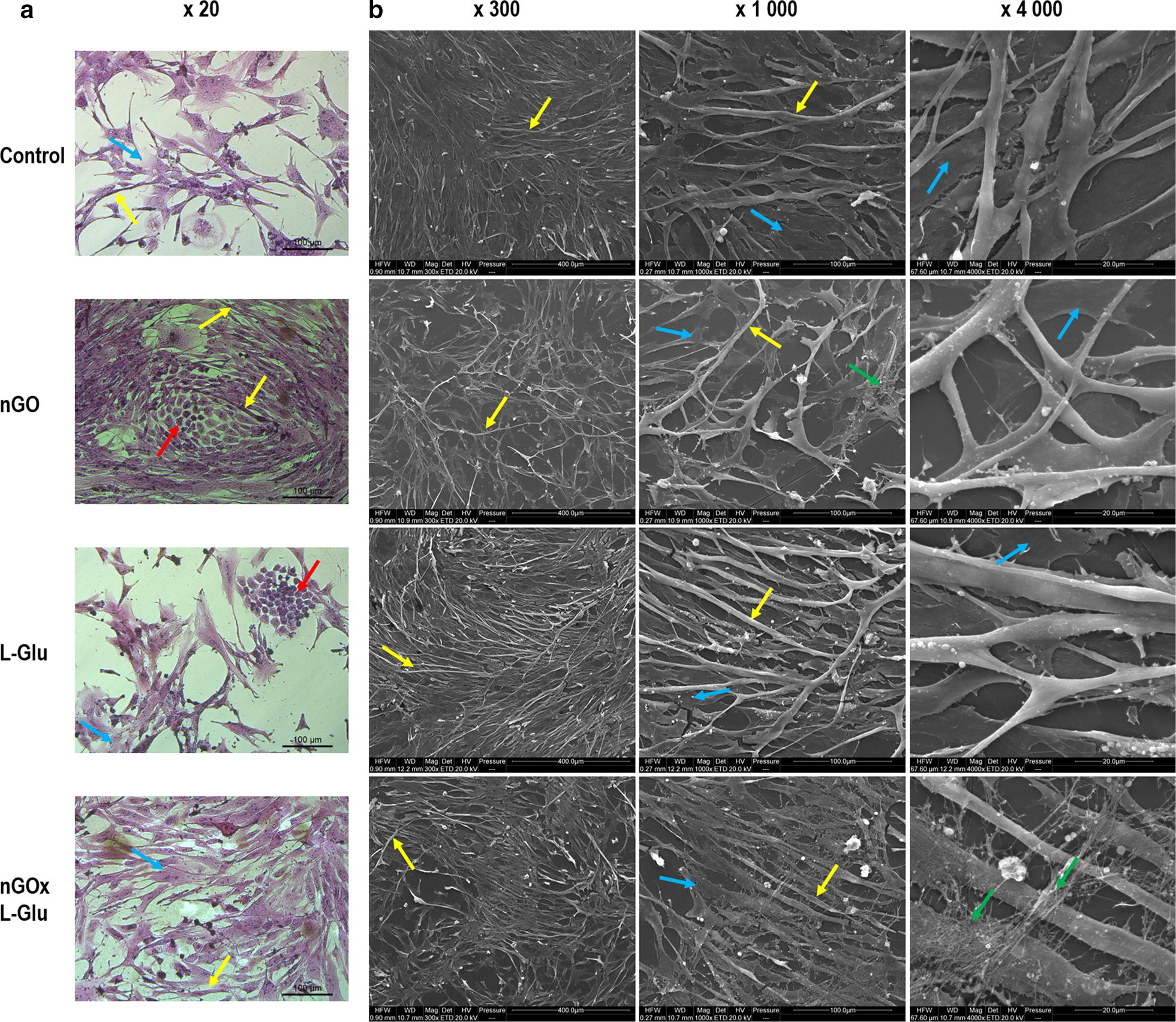
Fig. 4.

Cell morphology evaluated by optical microscopy (a) and scanning electron microscopy (b). a The images show cells cultured 5 days and hematoxylin and eosin stained; the red arrow indicates satellite cells, the yellow arrow indicates myofibres, the blue arrow indicates primary stromal cells; b the images show the control group cells cultured 5 days and the cells cultured 5 days on graphene oxide nanofilm (nGO), l-glutamine (L-Glu), and nGO with L-Glu supplementation (nGOxL-Glu); primary muscle fibres (yellow arrows), collagen fibres (green arrows), primary stromal cells (blue arrows)
