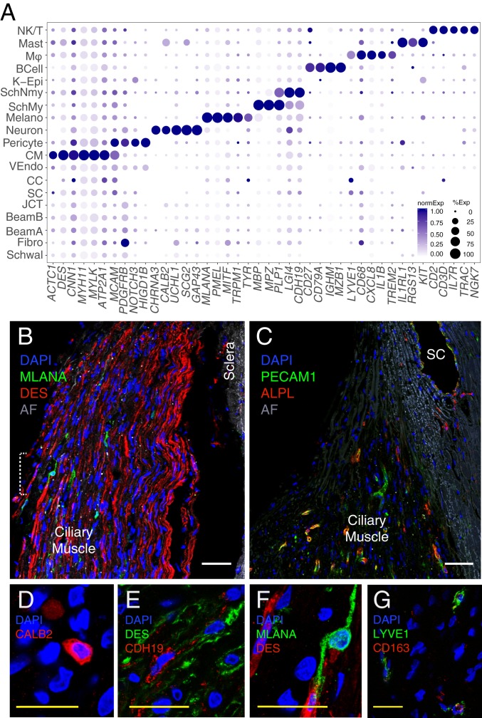
Fig. 4.
Cells of the human uveoscleral pathway. (A) Dot plot showing genes selectively expressed in cells of the uveoscleral outflow pathway. Fibro, fibroblast; K-Epi, corneal epithelium; Melano, melanocyte; MØ, macrophage; SchMy, myelinating Schwann cell; SchNmy, nonmyelinating Schwann cell; Schwal, Schwalbe line; VEndo, vascular endothelium. (B) Smooth muscle cells immunostained for DES (red) and melanocytes immunostained for MLANA (green) in CM. (C) Capillaries in the CM immunostained for PECAM1 (green) and ALPL (red). Occasional PECAM1+ALPL+ staining was also noted in SC, suggesting that this structure contains more than one cell type. (D) Immunostaining for CALB2 (red) highlights intrinsic neurons of the CM. (E) Immunostaining for LYVE1 (green) and CD27 (red) identifies macrophages in the TM. (F) Schwann cells in the CM immunostained for CDH19 (red) amid CM cells stained for DES (green). (G) Higher magnification of the area bracketed in B demonstrates an MLANA+ melanocyte (green). AF, autofluorescence; DAPI, 4′,6-diamidino-2-phenylindole (nuclear stain). (Scale bars: B and C, 50 µm; D–G, 25 µm.)

