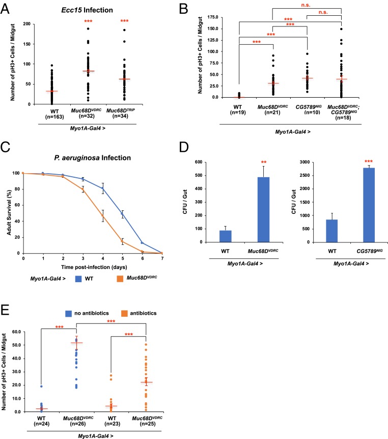
Fig. 5.
Intestinal stem cell phenotypes of Muc68D RNAi. (A) The average number of pH3+ cells in the posterior midguts expressing RNAi against Muc68D 24 h after Ecc15 oral infection. (B) The average number of pH3+ cells in the posterior midguts expressing RNAi against Muc68D, CG5789, and both Muc68D and CG5789. (C) Survival analysis of wild-type and Muc68D RNAi flies upon oral infection with P. aeruginosa. Error bars indicate SEM. (D) Internal bacterial load significantly increased in Muc68D RNAi and CG5789 RNAi flies. (E) The average number of pH3+ cells in the posterior midguts expressing RNAi against Muc68D with and without antibiotics treatment. (A, B, and E) “n” denotes the number of posterior midguts examined for each genotype. Error bars indicate SEM. **P < 0.05 and ***P < 0.001 (two-tailed t test). n.s., not significant.

