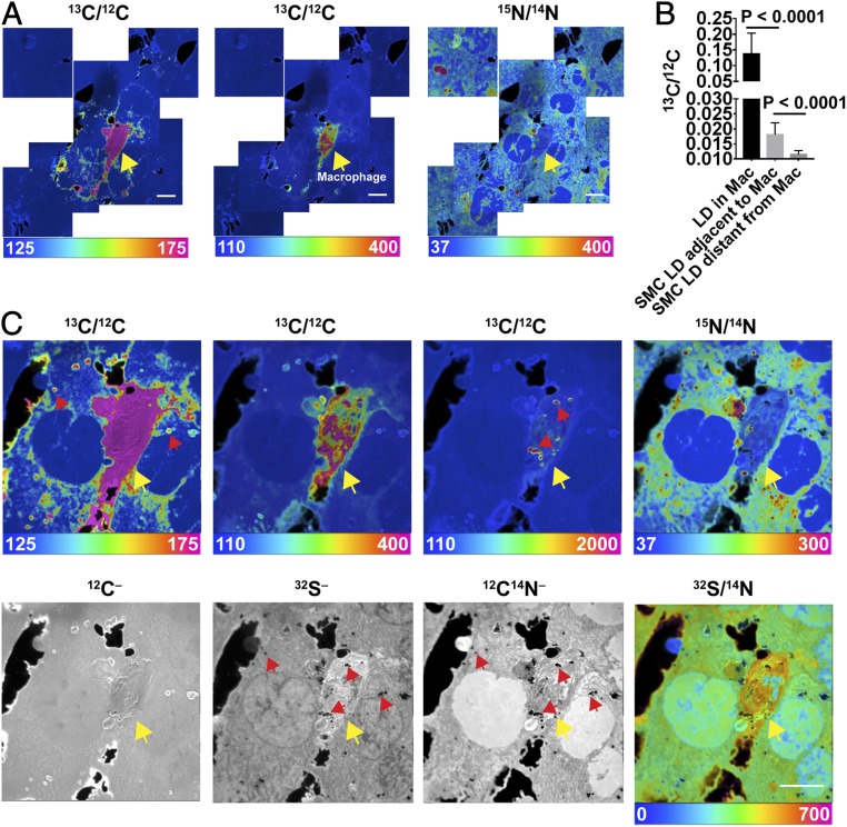
Fig. 1.
Transfer of [13C]cholesterol from [13C]cholesterol-loaded macrophages into adjacent SMCs. The SMCs had been grown for 21 d in medium containing [15N]choline. [13C]cholesterol-loaded macrophages were plated onto a ∼90 to 95% confluent monolayer of SMCs and incubated overnight at 37 °C in serum-free medium. NanoSIMS images were recorded from the interior of cells (note the presence of nuclei with negligible amounts of 15N enrichment). (A) A mosaic of 13C/12C and 15N/14N NanoSIMS images centered on a [13C]cholesterol-loaded macrophage. The macrophage is noted with a yellow arrow. The large black holes in the NanoSIMS images represent regions of the silicon wafer that were not covered by cells. (Scale bars: 10 μm.) Ratio scales were multiplied by 10,000. The natural abundance of 13C is 1.1%. 12C–, 32S–, and 12C14N– NanoSIMS images of the same field are shown in SI Appendix, Fig. S2. (B) 13C/12C ratio (mean ± SD) in macrophage (Mac) cytosolic lipid droplets (LD) (n = 23 LDs in five regions of 45 μm × 45 μm), LDs in SMCs immediately adjacent to the macrophage (n = 52 LDs in five regions of 45 μm × 45 μm), and LDs in more distant SMCs (n = 42 LDs in four regions of 45 μm × 45 μm). The cytosolic LDs in SMCs and macrophages were identified in the 32S– and 12C14N– NanoSIMS images (SI Appendix, Fig. S2). (C) High magnification image of the [13C]cholesterol-loaded macrophage shown in A. Ratio scales were multiplied by 10,000. The ratio scale of 125–175 shows the transfer of [13C]cholesterol from Macs to SMCs; the ratio scale of 110–400 shows [13C]cholesterol enrichment in Macs; and the ratio scale of 110–2,000 shows [13C]cholesterol enrichment in cytosolic LDs in Macs. Note that the macrophage is relatively enriched in 32S. Red arrows show a few examples of cytosolic LDs and yellow arrow points to the macrophage. (Scale bar: 10 μm.)

