Fig. 2.
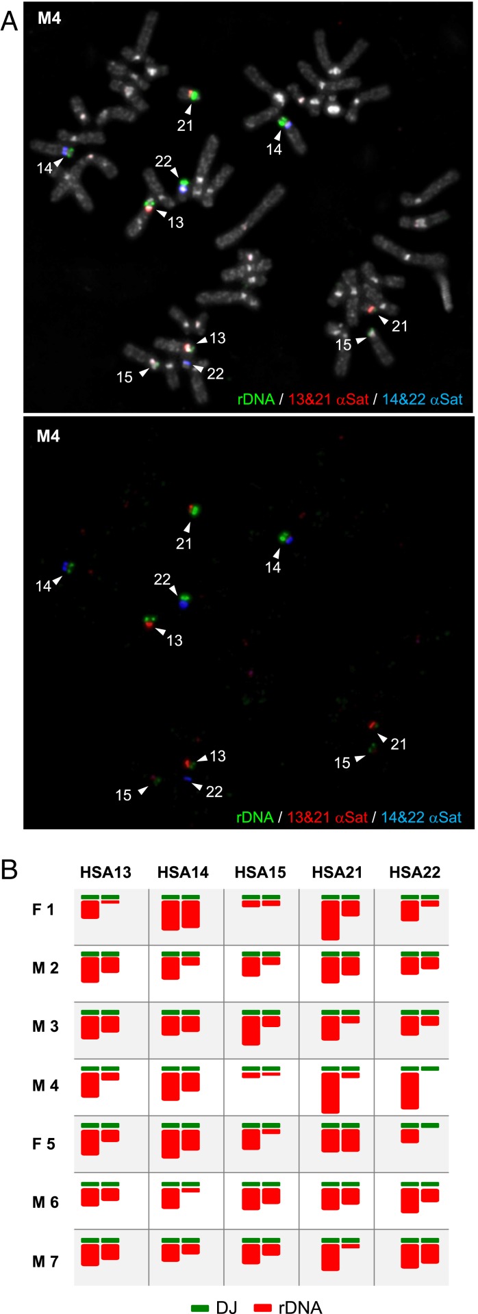
Chromosomal distribution of rDNA on metaphase spreads from human donors. (A) FISH performed on metaphase spreads prepared from a male donor, M4, using an rDNA (green) and α-satellite probes recognizing HSA13/21 (red) and HSA14/22 (far red, pseudocolored here in blue). Chromosomes were stained with DAPI (pseudocolored in gray). The identity of each acrocentric is indicated. (B) NOR ideograms showing the relative rDNA distribution in seven human donors, including M4 above (details in Materials and Methods).

