Fig. 5.
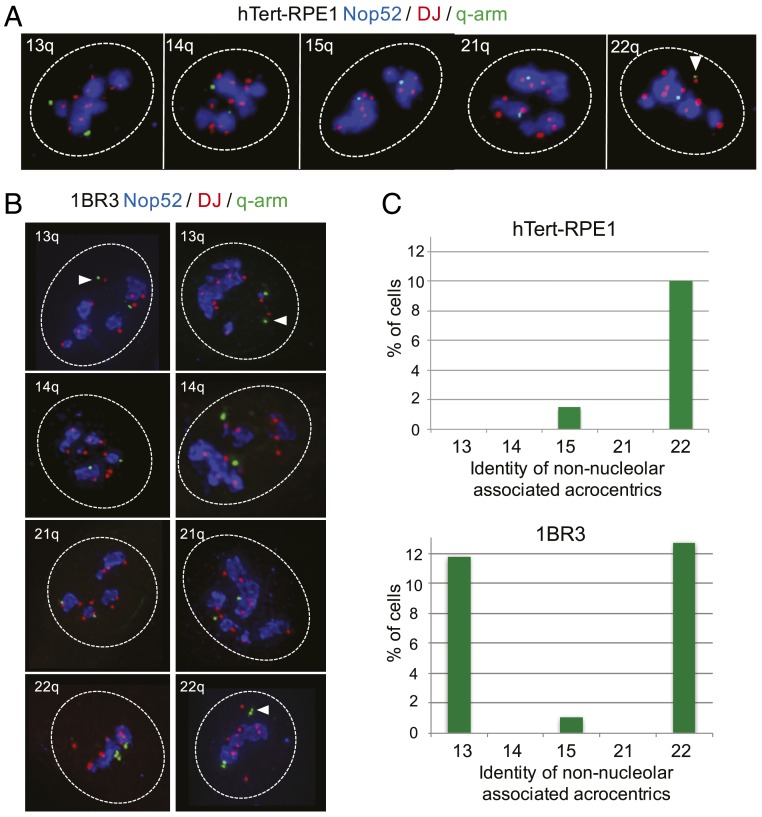
Identification of non–nucleolar-associating NORs. (A) 3D immuno-FISH performed on hTert-RPE1 cells. Nop52 antibodies (blue) used to visualize nucleoli were combined with DJ and q-arm BAC FISH probes (red and green, respectively). The identity of the q-arm probe is indicated in the upper right corner of each panel. Nuclear borders are indicated with dashed white lines, and the non–nucleolar-associated NOR from HSA22 is indicated by a white arrowhead. (B) 3D immuno-FISH performed on 1BR3 cells as above. (C) Quantitation of 3D immuno-FISH showing the percentage of cells in which the NOR from an identified acrocentric chromosome (shown below) is nonassociated with a nucleolus. Cell identities are shown above each bar chart. At least 100 cells were analyzed for each q-arm probe in each cell type.

