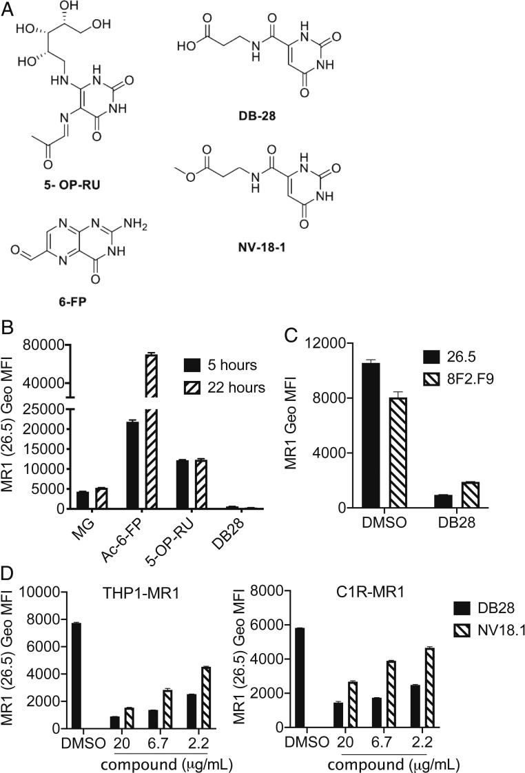
Fig. 2.
Characterization of DB28. (A) Chemical structures of the MR1 ligands used in this study: 5-OP-RU, 6-FP, DB28, and NV18.1. (B–D) DB28 and NV18.1 down-regulate MR1 from the cell surface. (B) THP1-MR1 cells were pulsed for 5 or 22 h with the indicated ligands (MG 50 μM, Ac-6-FP 1 μg/mL, 5-OP-RU 5 μg/mL, DB28 20 μg/mL) before staining with anti-MR1 (26.5) antibody. (C) THP1-MR1 cells pulsed overnight with DMSO or DB28 (20 μg/mL) were stained with the two indicated anti-MR1 antibodies. (D) THP1-MR1 (Left) or C1R-MR1 (Right) were pulsed overnight with the indicated concentrations of DB28 or NV18.1 before staining with anti-MR1 (26.5) antibody. Geo MFI ± SD of technical duplicates are plotted in each graph. Data representative of three experimental replicates.

