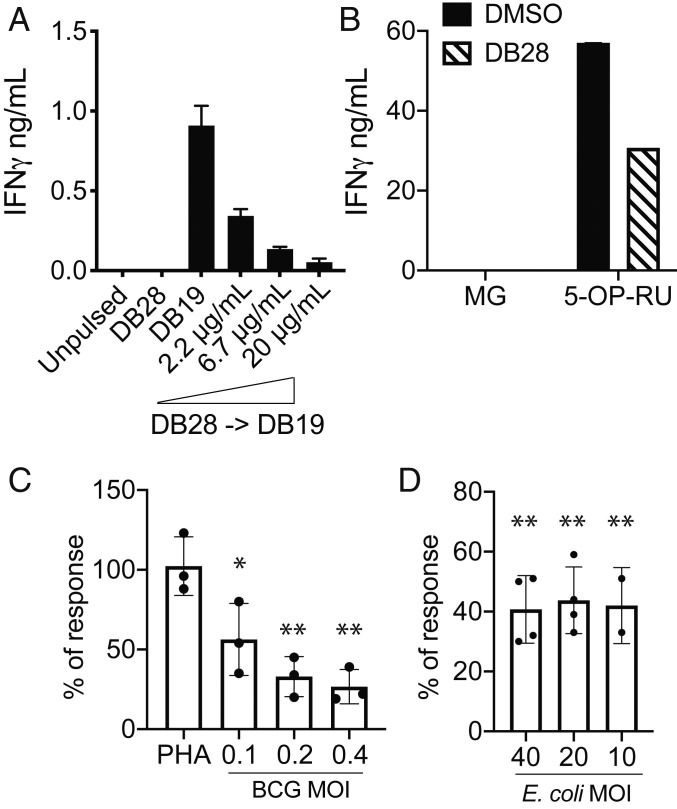
Fig. 7.
DB28 reduces the stimulatory activity of agonist-pulsed MR1-expressing antigen-presenting cells. (A and B) THP1-MR1 were pulsed with the indicated concentrations of DB28 2 h before pulsing with DB19 (20 μg/mL) (A) or 5-OP-RU (5 ng/mL) (B) and addition of MAIT cells. In B, DB28 was used at 20 μg/mL. IFN-γ (mean ± SD of technical duplicates) was measured in the supernatant collected after 16 h of stimulation. (C) MAIT cell stimulation by BEAS2B infected with bacillus Calmette-Guérin (BCG) at the indicated MOI, in the presence or absence of DB28 (20 μg/mL). Plotted is the percentage of response DB28/DMSO, where DMSO controls have been normalized to 100. n = 3 experimental replicates, each performed in technical duplicates (raw data for one donor shown in SI Appendix, Fig. S9B). Multiple t test, *P ≤ 0.05; **P ≤ 0.005. (D) MAIT cell stimulation in PBMC infected with the indicated MOI of E. coli in the presence or absence of DB28 (20 μg/mL). Plotted is the percentage of response DB28/DMSO. n = 4 experimental replicates (2 for MOI 10). (Raw data for one donor shown in SI Appendix, Fig. S9 C and D.) Multiple t test, **P ≤ 0.005.

