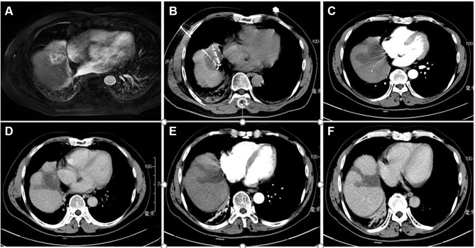
Figure 2.
Contrast-enhanced computed tomography (CT) revealed a HCC of 3.7 cm in maximum diameter adjacent to heart in segment 4 of a 57-year-old woman with liver cirrhosis caused by hepatitis B. (A) CT axial scan showed a high density round nodule adjacent to heart in segment 4 in arterial phase; (B) two CRA electrode probes were inserted into subcardiac HCC inside under CT guidance; (C) a clear ice-ball covered in HCC nodule is shown in arterial phase CT image after CRA; (D) low density CRA zone is shown in plain CT image after 3 months; (E) low density CRA zone is shown in arterial phase CT image after 3 months; (F) low density CRA zone is shown in delay phase CT image after 3 months.

