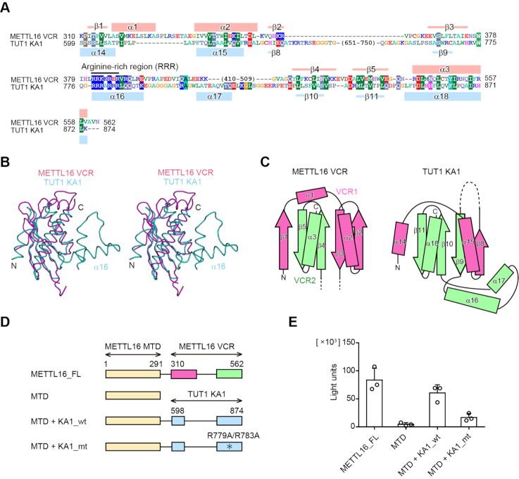
Figure 2.
The VCR is structurally and functionally equivalent to KA1 of TUT1. (A) Sequence alignments of the VCR of human METTL16 and the KA1 domain of human TUT1. The secondary structural elements of the VCR and KA1 domain are indicated above and below the alignments, respectively. (B) A stereo view of the superimposed structures of the VCR of METTL16 (purple) and the KA1 domain of TUT1 (cyan). (C) Topology diagrams of the VCR of METTL16 (left) and the KA1 domain of TUT1 (right). The N- and C-terminal halves are colored pink and green, respectively. (D) Schematic diagrams of METTL16 and its variants used for assays in (E). METTL16_FL: full-length METTL16, MTD: methyltransferase domain, MTD +KA1_wt: a chimeric protein of the N-terminal METTL16 MTD and the C-terminal TUT1 KA1, MTD+KA1_mt: mutant protein of MTD+KA1_wt. The asterisk indicates the R779A/R783A mutation. VCR1 and VCR2 of METTL16 are colored pink and green, respectively. (E) Methylation of U6 snRNA by METTL16_FL and its variants shown in (D), under the conditions in which 50 nM of U6 snRNA was used for the assays. Bars in the graphs are standard deviations (SD) of three independent experiments.

