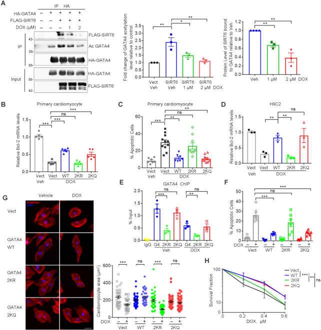
Figure 5.
Hypoacetylation of GATA4 diminishes cardio-protection against DOX. (A) Western blotting analysis of the levels of FLAG-SIRT6 protein and HA-GATA4 acetylation in the precipitated pool of HA-GATA4 with DOX treatment at 0, 1, 2 μM for 6 h (left). Percent level of acetylated GATA4 relative to vehicle/no FLAG-SIRT6 control (middle) and percent FLAG-SIRT6 bound to HA-GATA4 relative to vehicle control (right). Quantification was performed by ImageJ. n = 3. *P < 0.05. **P < 0.01. (B) qPCR analysis of Bcl-2 mRNA levels in primary mouse neonatal cardiomyocytes ectopically expressing GATA4 WT, K328/330R (2KR) and K328/330Q (2KQ) upon exposure to DOX (1 μM, 6 h). ***P < 0.001, ‘ns’ represents no significance. (C) Percent TUNEL positively stained primary cardiomyocytes after treatment with DOX (1 μM) for 24 h. **P < 0.01, ***P < 0.001, ‘ns’ indicates no significance. (D) H9C2 cells stably expressing GATA4 WT, K328/330R (2KR) and K328/330Q (2KQ) were exposed to DOX (1 μM, 6 h). The mRNA transcripts of Bcl-2 were evaluated by qPCR. **P < 0.01, ‘ns’ represents no significance. 18srRNA was used as a reference gene. (E) ChIP analysis was performed in the stably transfected H9C2 cells by using anti-GATA4 antibody in the presence/absence of DOX. GATA4 enrichment on the Bcl-2 promoter region was measured by qPCR. ***P < 0.001, **P < 0.01, ‘ns’ represents no significance. (F, G) Stably transfected H9C2 cells were exposed to DOX (1 μM) for 24 h and stained with TUNEL or TRITC-phalloidin. Images of the stained cells were captured. The quantification of the number of positively TUNEL stained H9C2 cells (F). Cell area was measured and quantified (G). Scale bar, 20 μm. ***P < 0.001, ‘ns’ represents no significance. (H) Cell colony formation showing the survival fraction of H9C2 cells after the indicated doses of DOX for 1 h. The plates (n = 3/group) were stained with crystal violet and the colony numbers were counted. **P < 0.01, ***P < 0.001, ‘ns’ represents no significance.

