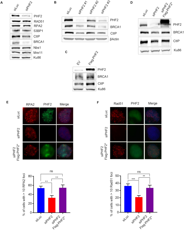
Figure 3.
PHF2 regulates HR by modulating CtIP and BRCA1 levels. (A) U2OS cells were depleted for Luc or PHF2 by siRNA. Forty eight hours later, the cells were lysed and extracts were analysed by western blot with the indicated antibodies. (B) U2OS cells were depleted for PHF2 using three different siRNA oligonucleotides and subsequently analysed by western blot using the indicated antibodies. (C) U2OS cells were transfected with empty vector (EV) or a Flag-PHF2 expression vector, followed by western blot analysis with the indicated antibodies. (D) U2OS cells were depleted for Luc or PHF2 by siRNA and 24 h later transfected with EV or siRNA-resistant Flag-PHF2 (Flag-PHF2*). The day after, extracts were prepared and analysed by western blot with the indicated antibodies. (E) U2OS cells were depleted for PHF2 and transfected with Flag-PHF2* the day after. One day later, cells were treated with IR (3 Gy) and fixed for IF after 1 h. RPA2 focus formation of Flag-positive cells was analysed by immunofluorescence. Top panel: representative images. Bottom panel: quantification of three independent experiments, counting at least 50 cells each. (F) U2OS cells were depleted for PHF2 and transfected with Flag-PHF2* the day after. One day later, cells were treated with IR (3 Gy) and fixed for IF after 4 h. Rad51 focus formation of Flag-positive cells was analysed as in (E).

