Figure 2.
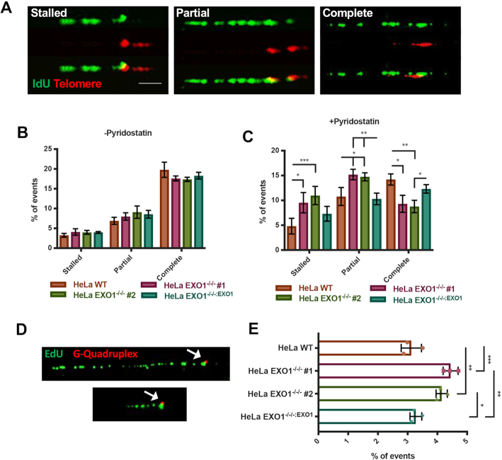
EXO1 deficiency leads to telomere replication defects under G4-stabilizing conditions. (A) Representative images of telomere replication events. Replication tracts that extend to the end of the telomere were scored as complete, tracts that are partially replicated were scored as partial and tracts at the beginning of the telomere sequence were denoted as stalled. Scale bar = 10 kb. (B) Quantification of telomere replication events in EXO1 WT, EXO1−/−/− #1 and #2, and EXO1−/−/−:EXO1; n = 200 from three independent experiments. (C) Quantification of telomere replication events in EXO1 WT, EXO1−/−/− #1 and #2, and EXO1−/−/−:EXO1 with 7.5 μM pyridostatin for 5 h prior to and through labeling. Statistical significance was determined by one-way ANOVA analysis followed by Tukey's post-hoc test; n = 200. (D) Representative images of native replication tracts (green) stalled at a G4 (red). (E) Quantification of replication forks stalled at G4s with 7.5 μM pyridostatin for 5 h prior to and through labeling. Significance was determined by one-way ANOVA analysis followed by Tukey's post-hoc test; n = 600 from three independent experiments.

