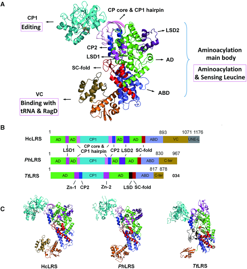
Figure 1.
The overall structure of hcLRS. (A) Overall structure of hcLRS shown in cartoon. The Rossmann-fold domain (AD, green), the CP core & CP1 hairpin (pink), the CP1 editing domain (cyan), the CP2 domain (blue), the eukaryotic leucyl-specific domains 1 (magenta) and 2 (violet-purple), the SC-fold domain (red), the α-helix bundle domain (slate) and the VC domain (orange and sand); (B) A diagram shown the domains and motifs of hcLRS, PhLRS and TtLRS; (C) Structural comparison of hcLRS, PhLRS and TtLRS shown in cartoon from a same view.

