Figure 2.
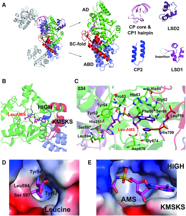
Aminoacylation active site of hcLRS and the binding details of LeuAMS. (A) The cartoon representation of aminoacylation main body of hcLRS, each inserted motifs are dissected from the structure for a clear view. (B) Shows HIGH motif (His 60 to His 63, blue) and KMSKS motif (Lys716 to Ser720, yellow), and the LeuAMS is shown in stick; (C) Binding details of the LeuAMS (stick representation) in the aminoacylation active site, residues within 4 Å are labeled. The binding pocket (electrostatics surface representation) of LeuAMS are shown separately for the Leucine moiety binding pocket (D) and AMS moiety binding pocket (E).

