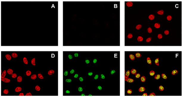
Figure 10.
Evaluation of cell entry by confocal microscopy. Images of (A) untreated TZM-bl cells, TZM-Bl cells treated with 5 × 10⁻⁵ M of (B) NDI-PNA 6, (C) NDI 6 or (D–F) NDI-PNA 8. The –compounds were incubated with the cells for 2 h before cell fixation in 2% PFA. Nuclear staining was obtained with Nuclear Green LCS1. For NDI/conjugates (red channel) images (A-D) were visualized at 561 nm excitation wavelength and 570–620 nm emission range; for cell nuclei (green channel) (E) 488 nm excitation wavelength and 500–550 nm emission range were applied. (F) Overlap of panels (D) and (E).

