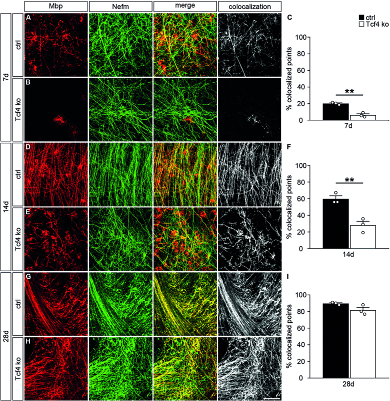
Figure 4.
Impaired oligodendroglial differentiation in organotypic slice cultures of Tcf4ko mice. (A, B, D, E, G, H) Immunohistochemical stainings of organotypic slices from forebrain of control (ctrl) (A, D, G) and Tcf4ko (B, E, H) newborn mice with antibodies directed against Mbp (red) and Nefm (green) after seven days (7d) (A, B), 14 days (14d) (D, E) and 28 days (28d) (G, H) in culture. Single stainings are shown (two left panels) as well as their merge (third panel) and the colocalizing signal (white, right panel). Scale bar: 100 μm. (C, F, I) From these and similar stainings, quantifications were performed to determine the relative area with colocalized Mbp and Nefm signals using the Colocalization Threshold plugin of ImageJ (n = 3 slices from separate animals per genotype, counting at least 0.15 mm2 per slice). Differences to controls were statistically significant after 7 and 14 days in culture as determined by two-tailed Student's t test (**P ≤ 0.01).

