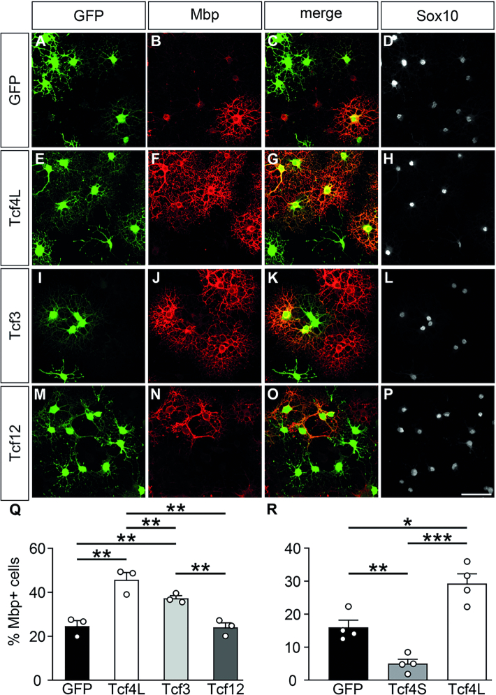
Figure 5.
Influence of increased expression of class I bHLH proteins on oligodendrocyte differentiation in vitro. (A–P) Immunocytochemical stainings of rat oligodendroglial cells with antibodies directed against Gfp (green, A, E, I, M), Mbp (red, B, F, J, N) and Sox10 (white, D, H, L, P). Cells were kept for 6 days in differentiating conditions after transduction with control (A–D), Tcf4L- (E–H), Tcf3- (I–L) and Tcf-12 (M–P) expressing retrovirus. Single stainings are shown as well as their merge (C, G, K, O) as indicated above the panels. Scale bar: 50 μm. (Q) Quantifications were performed to determine the fraction of Mbp-expressing cells among all transduced cells (n = 3 cultures from separate transductions, counting at least four representative visual fields per transduction). (R) In a similar manner, quantifications were performed to determine the fraction of Mbp-expressing cells among cells transfected with expression plasmids for GFP alone or in combination with Tcf4L and Tcf4S isoforms (n = 4 separately transfected cultures, counting at least four representative visual fields per transfection). Differences to controls were statistically significant as determined by one way ANOVA with Bonferroni correction (*P ≤ 0.05; **P ≤ 0.01; ***P ≤ 0.001).

