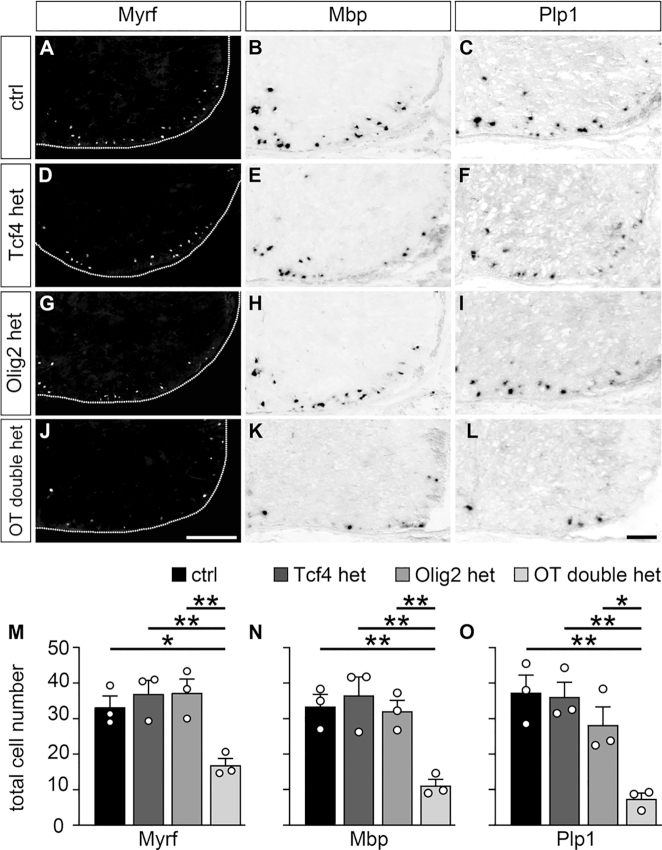
Figure 9.
Impaired oligodendroglial differentiation in OT double het mice. (A–L) Immunohistochemical stainings with antibodies directed against Myrf (A, D, G, J) and in situ hybridizations with probes specific for Mbp (B, E, H, K) and Plp1 (C, F, I, L) were carried out on spinal cord sections from wildtype control (ctrl, A–C), Tcf4 het (D–F), Olig2 het (G–I) and OT double het (J–L) mice at E18.5. The right ventral horn is shown. For immunohistochemical stainings, the spinal cord was placed on a black background and demarcated by a stippled line. Scale bar: 100 μm. (M–O) From these and similar experiments, quantifications were performed to determine the absolute mean number of marker-positive cells ± SEM per thoracic spinal cord section (n = 3 mice for each genotype, counting three separate sections). Differences to controls were statistically significant as determined by one way ANOVA with Bonferroni correction (*P ≤ 0.05; **P ≤ 0.01).

