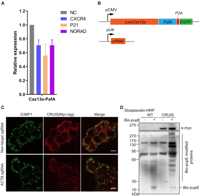
Figure 2.
Testing the activity of CRUIS. (A) HEK239T cells were co-transfected with LwaCas13a-PafA and sgRNA expression plasmid to detect the mRNA expression level of the target gene after 24 hours; non-target sgRNA was used as the negative control (n = 3, mean ± S.E.M). (B) Plasmids used in this assay. (C), Representative immunofluorescence images of 293T-CRUIS cells treated with 100 mM sodium malonate (scale bar 10μm). Stress granules are indicated by G3BP1 staining. (D) Testing the proximity label activity of CRUIS.

