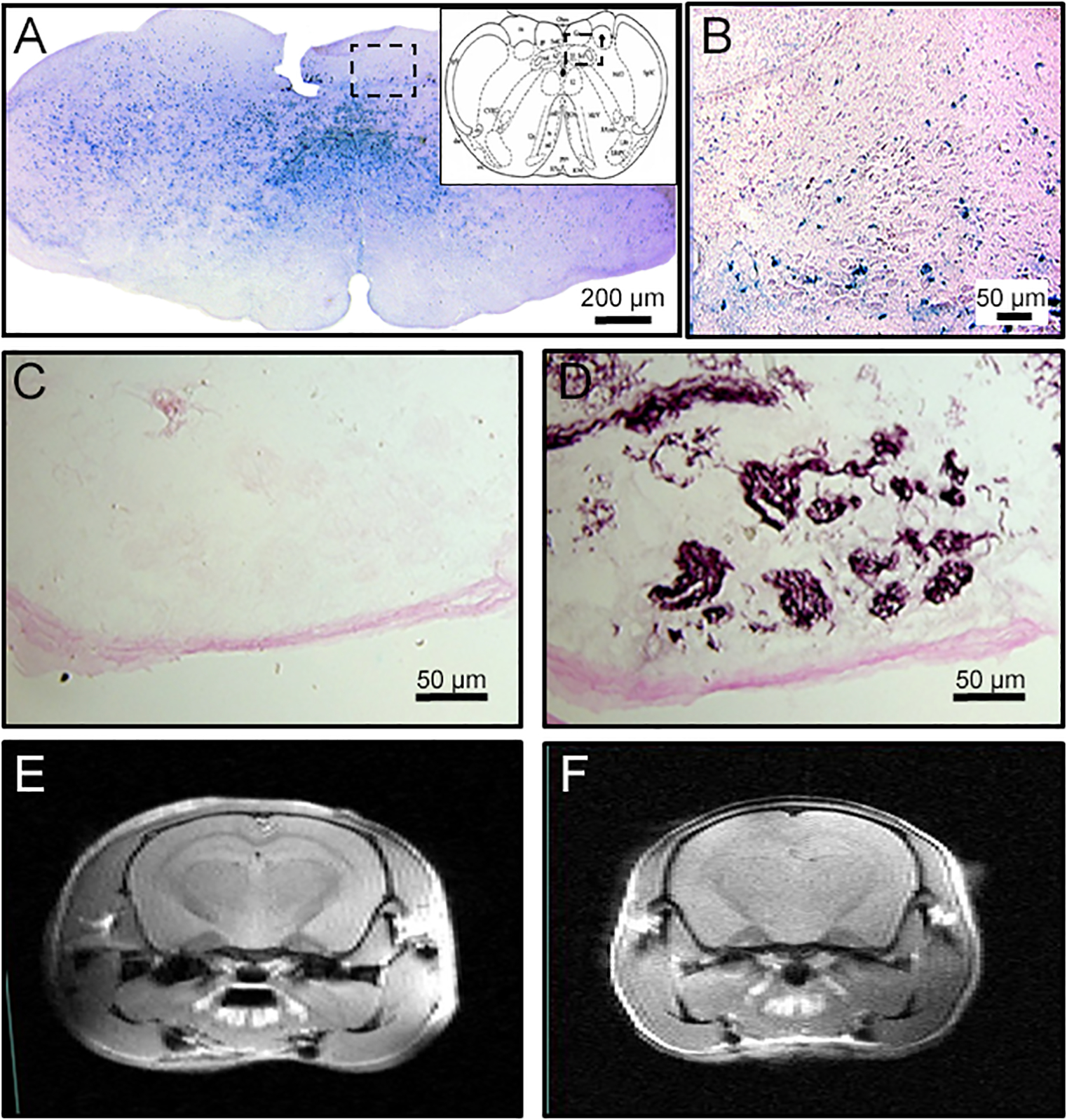
Figure 1.

Constitutive deletion of HIF-1α in the central but not peripheral nervous system using the Cre-LoxP strategy. A. Cre is expressed throughout the brainstem as demonstrated by Lac-Z staining (blue) in reporter mice, including the NTS (B, magnified set from A). However, Lac-Z is not observed in in the carotid body (C) which has normal chemosensitive glomus cells as shown with tyrosine hydroxylase staining in a consecutive section (D). MRI images show normal brain structure and ventricles in wild-type (CNS-HIF-1α+/+, E) or CNS-HIF-1α−/− null mice (F).
