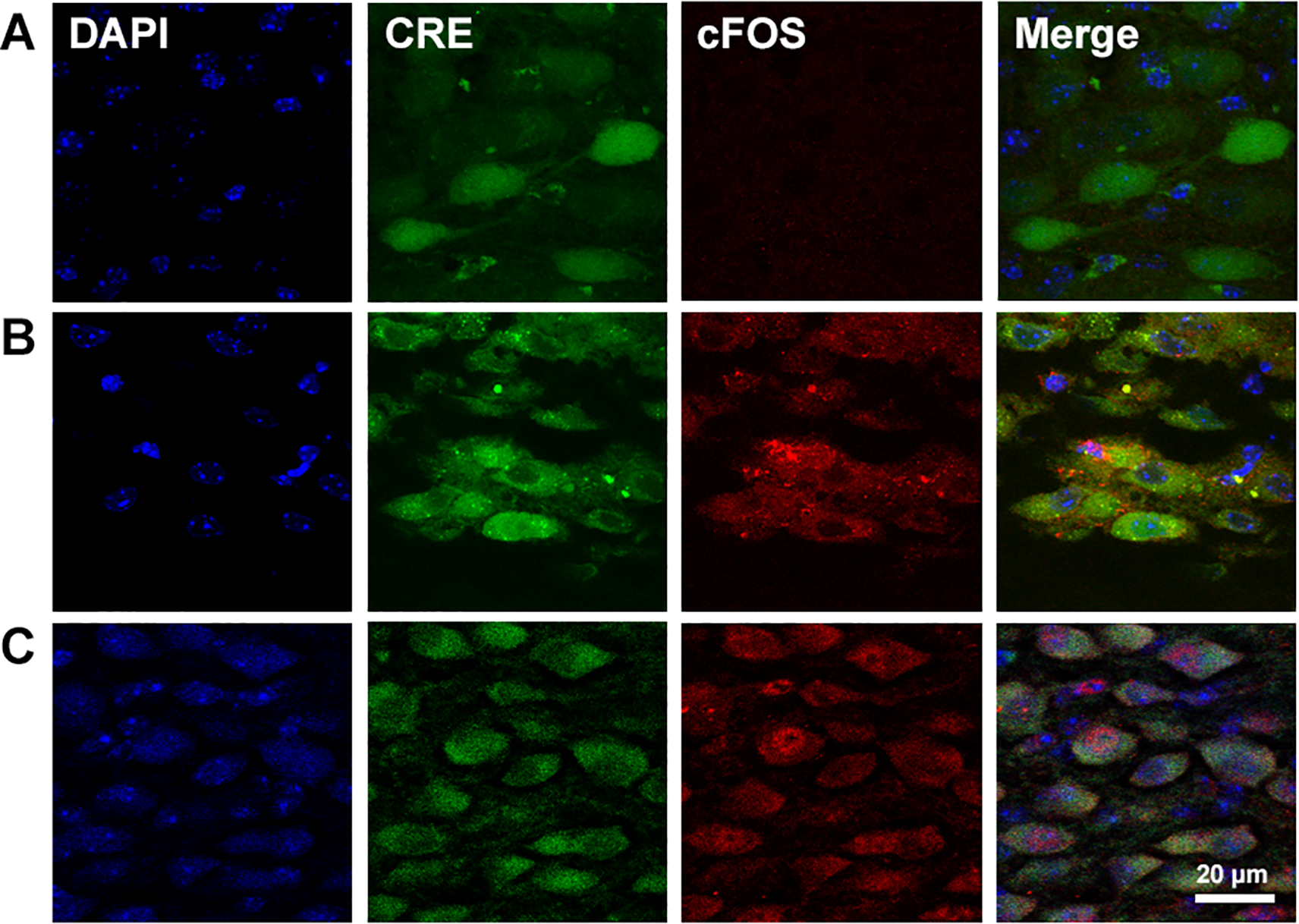
Figure 6.

Micro-photographs showing neurons in the NTS infected with AAV in a mouse exposed to control normoxic conditions (A), to 3 hours of hypoxia (B) or 7 days of CH (C). Cellular nucleus were stained with DAPI (blue). Expression of GFP-Cre recombinase (Cre, green) allows to observe AAV-infected neurons. Expression of cFOS (red) was observed in cytoplasm of AAV-infected neurons after 3 hours of hypoxia (B) and in the cell nucleus in the CH mouse (C), but not in the control mouse (A). The overlapped image (merge) shows that AAV is infecting neurons that are activated by 3 hours of hypoxia.
