Fig. 1.
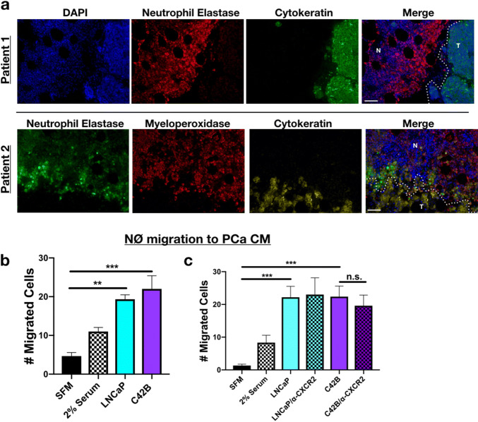
PCa recruitment of neutrophils. a Representative immunofluorescence (IF) of PCa and neutrophils in bone marrow of BM-PCa patients. Top: Patient 1—neutrophil elastase (NE; red) and epithelial marker, pan-cytokeratin (green), and nuclear marker, DAPI (blue); Bottom: Patient 2—neutrophil elastase (green), myeloperoxidase (red), cytokeratin (gold), DAPI (blue). “N” denotes normal bone marrow, “T” denotes a region of tumor in bone. Size bar = 50 μm. b Boyden chamber migration assay and shows number of neutrophils that migrated through the Boyden membrane into the lower chamber. Neutrophils were allowed to migrate toward specific conditions, for 1 h: serum-free media, serum containing 2% FBS, serum-free LNCaP conditioned media (CM), and serum-free C42B CM. c Neutrophil migration assay toward PCa media supplemented with an antibody to mouse CXCR2 (50 nM). Asterisks denote statistical significance (**p < 0.01, ***p < 0.001)

