Fig. 6.
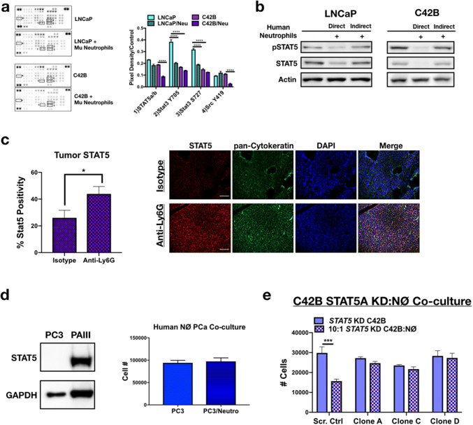
Neutrophils induce PCa death via inhibition of STAT5. a Phospho-kinase protein array of LNCaP and C42B cells cultured alone or with mouse bone marrow-derived neutrophils (denoted as Mu neutrophils). Boxes correspond to the genes labeled in. Three samples of each condition were pooled, and 100 μg of protein loaded onto the array. Proteins are displayed in duplicate. Average dot pixel density was averaged per protein target, background density subtracted, and normalized to the reference control per blot. Densitometry analysis was performed using Image J software. Graph represents pixel density quantitation of pSTAT5, pSTAT3, and pSrc. Asterisks denote statistical significance (****p < 0.0001). b STAT5 western blot of whole-cell PCa (30 μg protein) lysates from direct and indirect co-cultures of human neutrophils with LNCaP (left) and C42B (right) cells. For indirect co-cultures, modified Boyden chamber assay was utilized. c Immunofluorescence quantitation is the percentage of STAT5-positive C42B per total cell number (left) and representative images (right) of Total STAT5 (red) in C42B bone metastases (n = 5). C42B tumor cells in bone are denoted by cytokeratin (green), and nuclei are stained by DAPI (blue). Size bar = 20 μm; asterisk denotes statistical significance (*p < 0.05). d Left, Western blot analysis of Stat5 in PC3M compared to PAIII. Right, PC3M cell counts 24 h after direct co-culture with human bone marrow neutrophils. e Direct co-culture of bone-derived mouse neutrophils and C42B cells expressing: a non-targeted scrambled sequence shRNA (Scr. Ctrl) or STAT5A shRNA plasmids (denoted STAT5 knockdown (KD) clones A, C, and D). Graph shows the total number of C42B cells 24 h after co-culture measured by Trypan Blue Exclusion assay. Asterisk denotes statistical significance (***p < 0.001)

